
We are happy to announce the winners of the (MC)2 annual image contest. The contest was judged by center staff and featured two categories: (1) Transmission Electron Microscopy and (2) Scanning Electron Microscopy/X-ray Microscopy/Other. Thank you to everyone who participated for the high number of quality submissions, representing diverse research subjects and techniques. We still hope to feature some of the images that were not selected for prizes in other ways in the future.
Transmission Electron Microscopy
1st Place
Nishkarsh Agarwal, “Glacial Microbloom: Defect Anomaly at Cryo-Temperatures,” Hovden Research Group
Acquired on the TFS Talos
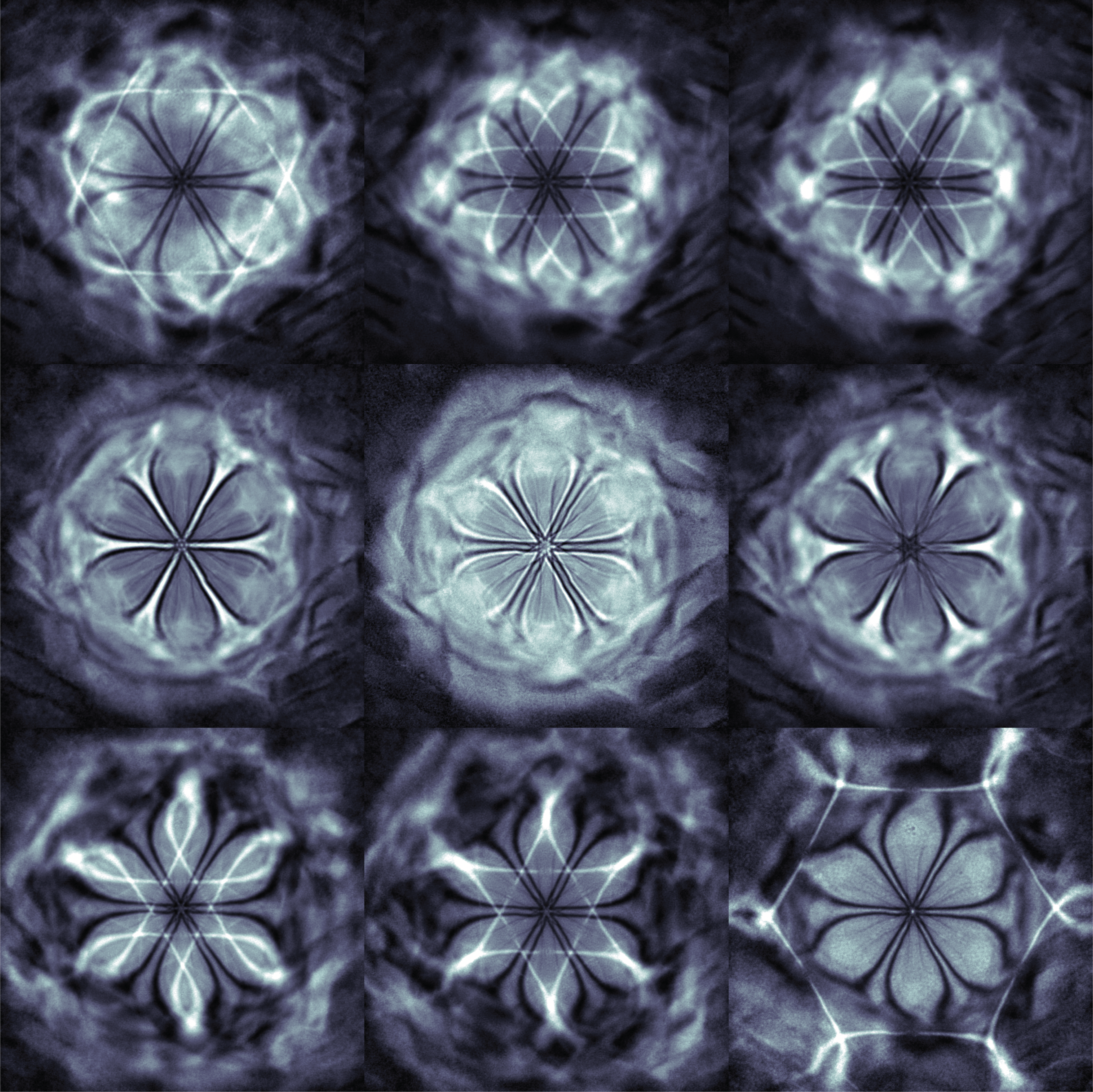
Nishkarsh Agarwal, a doctoral student in materials science and engineering, captured this series of images when searching for signatures of magnetic transitions in iron phosphorus trisulfide (FePS3) – a transition metal dichalcogenide van der Waals material that has magnetic phase transitions at low temperatures.
“Generally, these magnetic phases are difficult to probe through TEM, but we were hoping to observe this magnetic transition in real time to better understand the underlying mechanism and physics of the system,” said Agarwal.
During an in situ TEM at -176°C, a microscale distortion bubble in an ultrathin flake of FePS3 revealed the 6-fold symmetry of the underlying atomic structure. The non-monotonic CTF of TEM combined with bend contours from the flake’s pinning point and Kikuchi lines from the corresponding atomic planes created the flower-like image.
The SAEDs in the vicinity of the distortion have Bragg peaks oriented in the same direction as the “flower petals,” confirming the underlying crystal structure caused the 6-fold symmetry.
“When imaging, I was not initially surprised as pinning defects are common in thin 2D materials,” said Agarwal. “However, out of curiosity, I took a magnified picture of one such defect, and I saw these intricate features which intrigued me. I have never seen such clear 6-fold patterns from a defect. These features changed based on the TEM imaging conditions, and I took multiple series of images.”
When he’s not in the lab, Agarwal enjoys running, spending time with friends, and perusing YouTube videos.
Nishkarsh Agarwal captured “Glacial Microbloom” while searching for signatures of magnetic transitions in the iron phosphorus trisulfide (FePS3)
2nd Place
Arkajit Ghosh, “Fairy Lights of Atoms,” Misra Research Group
Acquired on the TFS Spectra

Arkajit Ghosh, a doctoral student in materials science and engineering, took this winning image while investigating the microstructure of rapid solidified aluminum-germanium eutectics.
Ghosh discovered a new aluminum-germanium hexagonal close packed (HCP) phase using high-angle annular dark-field imaging (HAADF) STEM technique and atomic scale Energy Dispersive X-ray (EDX) analysis. In the image, bright dots correspond to pure germanium columns and dark spots correspond to overlapped aluminum and germanium atomic columns.
“I was happy indeed after taking these images once I realized that it was possibly a new phase,” said Ghosh.
When he’s not conducting STEM imaging techniques, Ghosh enjoys photography and cooking.
Arkajit Ghosh took “Fairy Lights of Atoms” while investigating the microstructure of rapid solidified aluminum-germanium eutectics
3rd Place
Yanan Wang, “Nano-Twigs,” Kotov Research Group
Acquired on the TFS Talos
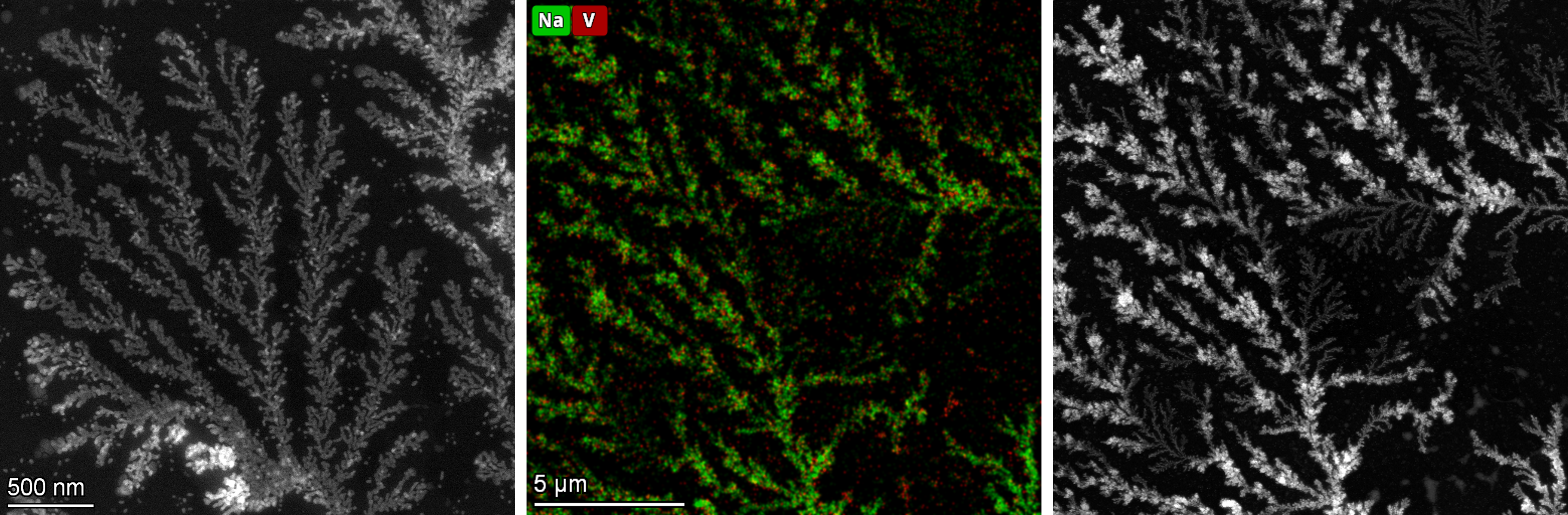
Yanan Wang, a postdoctoral visiting scholar in chemical engineering, captured these delicate branching patterns during Transition Electron Microscopy (TEM) characterization of vanadium oxide nanoparticles dissolved and stabilized under sodium buffer solution.
“Vanadium oxide nanoparticles are very dynamic, and the optical properties can be affected by the solution’s pH. We set samples with buffers of varying pH levels before imaging to figure out if they create distinguishable structures,” said Wang.
Wang enjoys cooking and is learning the Argentine tango.
Yanan Wang captured “Nano-Twigs” during TEM characterization of vanadium oxide nanoparticles
SEM / X-ray Microscopy / Other
1st Place
Zane Zhang, “Dried Riverbed,” Pena-Francesch Research Group
Acquired on the TFS Nova
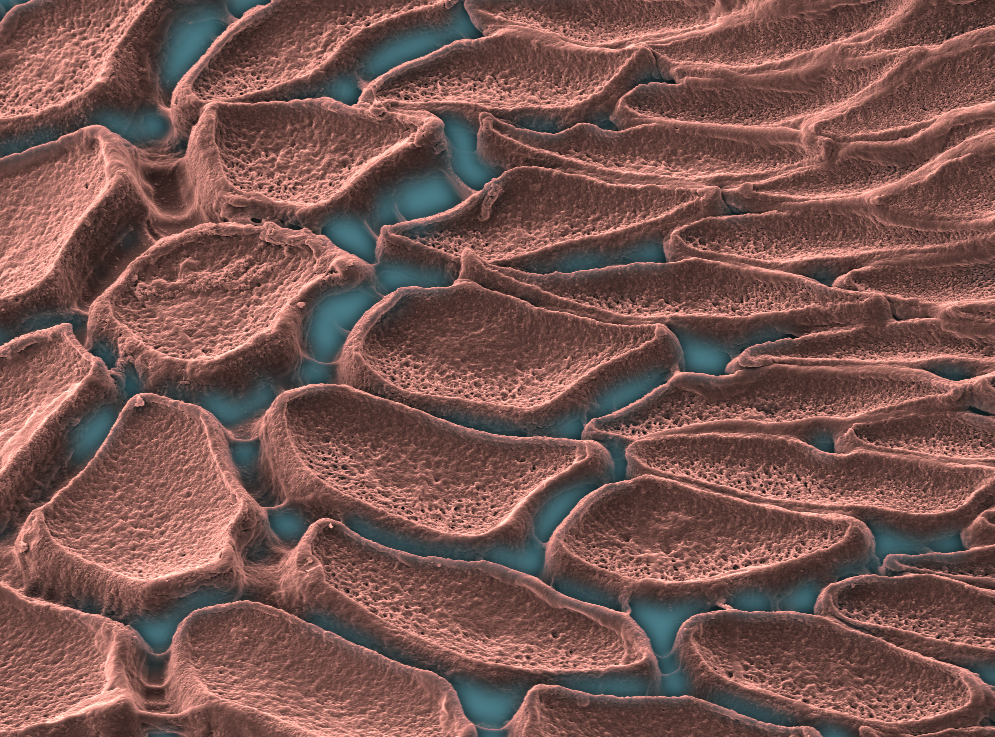
When investigating a self-healing protein derived from the “ring teeth” that encircle the suction cups at the ends of squid’s tentacles, Zane Zhang found the surrounding tissue resembles a dried riverbed through the lens of the TFS Nova Scanning Electron Microscope.
“While moving the stage, I vaguely saw this tissue attached to the teeth with these exciting patterns. I tuned the focus on this tissue and took the image,” said Zhang, a doctoral student in materials science and engineering.
The Pena-Francesch Research Group is developing robust, biodegradable materials and microrobots that incorporate this self-healing protein. Their module-based, rearrangeable microrobots are controlled by magnetic fields. These microrobots have the potential to aid in minimally invasive surgery or surgical tasks that require tiny, precise movements.
When he’s not leading a squid teeth photoshoot, Zhang enjoys taking road trips.

Zane Zhang discovered captivating patterns when imaging squid “ring teeth” that encircle the suction cups
2nd Place
Aramanda Shanmukha Kiran, “Eutectic Microstructure in 3D,” Shahani Research Group
Acquired on the Zeiss Xradia Versa 520
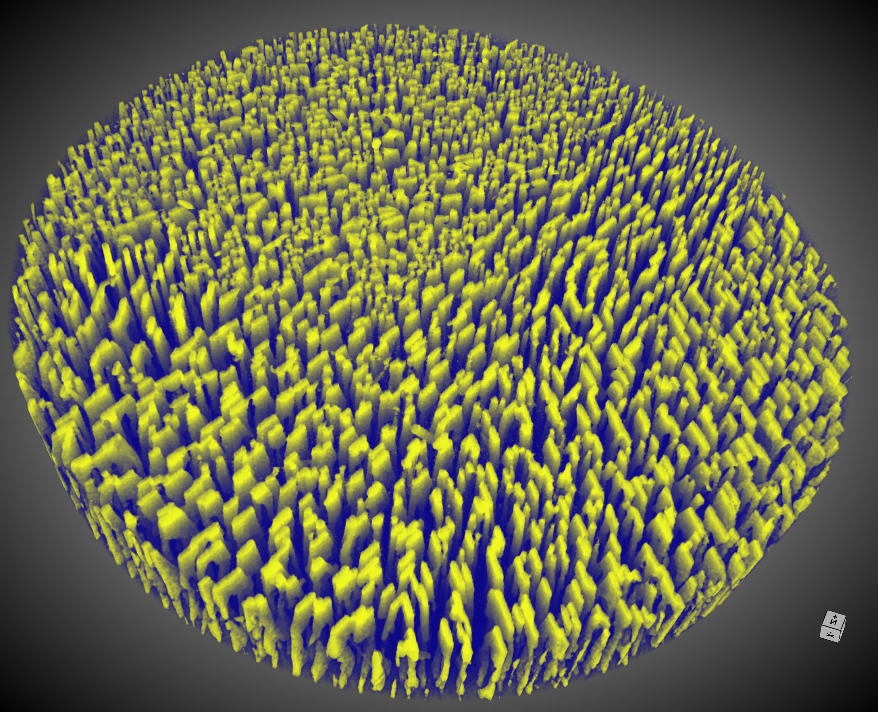
Aramanda Shanmukha Kiran, a postdoctoral research fellow in materials science and engineering, captured this school spirited image when exploring pattern formation during solidification of an aluminum-nickel eutectic alloy.
X-ray microtomography allowed observation of the intricate three-dimensional morphology. Studying pattern formation during eutectic solidification, particularly in systems with a low percentage of faceted phases, contributes to the development of distinctive microstructures.
“When I came across this image, I was genuinely thrilled and fascinated by the exotic microstructure it revealed. The level of detail and the unexpected beauty within the microstructure left me in awe and further motivated my enthusiasm for exploring and understanding these intricate formations,” said Kiran.
Beyond microstructures, Kiran enjoys the art and science of cooking and playing chess and badminton.
Aramanda Shanmukha Kiran revealed the intricate microstructure of an aluminum-nickel eutectic using X-ray microtomography
3rd Place
Arit Patra, “Vortice-Shaped Flowers in the Nanoscale,” Lahann Research Group
Acquired on the Helios 650

Arit Patra, a doctoral student in chemical engineering, came across this flower-like assembly of nanofiber arrays when investigating polymerization into topological point defects inherent to thin liquid crystalline films.
“I was astonished to see the perfect defect structure with proper contrast. The image depicts theory with clear accuracy,” said Patra.
Patra used the liquid crystalline material’s defects to guide chemical vapor deposition (CVD) polymerization. The resulting material allowed exploration of the optical characteristics.
When he’s not in the lab, Patra enjoys biking and reading.

Arit Patra came across flower-like vortices of nanofiber arrays when using a liquid crystalline material’s defects to guide chemical vapor deposition polymerization