In the past, we have brought portable scanning electron microscopes to schools and museums for interactive science demos. We can also work with you to organize a field trip to our lab so students can have a hands-on experience learning about and operating our scanning electron microscopes. If you are interested in planning any activities for your class, let us know!
Contact: Bobby Kerns ([email protected])
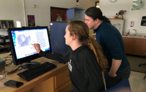
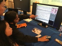
Dexter High School, MI (January 2018):
“Anytime we can offer our students a real world laboratory experience it enhances the experience of our students, increasing their drive to further their studies in the sciences upon graduation.”
—Beau Kimmey, Science Teacher
The SEM was located at Dexter High School for over a month from mid-December to late January. During that time lead science teacher Daniel “Beau” Kimmey and his colleagues used it in a number of classes. John presented two half day demonstrations of the instrument to Beau, two other science teachers in the school, and also to groups of students who were shown the applications of SEM to metals, semiconductors, biomaterials, insects, leaves, solder, shark skin, butterfly wings, etc.
“Through the use of the electron microscope I have seen first hand the engagement and excitement our students have where they can use a hands on approach rather than a passive style of learning.”
—Beau Kimmey
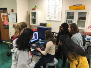
Northville High School, MI (November 2017):
The Hitachi TM3030 SEM was located at Northville High School for over a month from mid-October to late November. During that time lead science teacher Richard Cole and his colleagues used it in a wide variety of classes. John Mansfield presented several half day demonstrations of the instrument to Richard, other science teachers, and students.
“The microscope helped students make the idea of atoms which can be an abstraction into something more concrete. It gave the students a better understanding of the atomic scale. Many students came before and after hours to study items of their interest. The students felt like real scientists!
—Richard Cole, Science Teacher
Butterfly Festival – University of Michigan Museum of Natural History (May 2017)
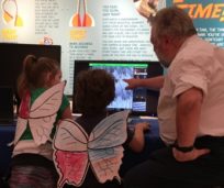
The Michigan Center for Materials Characterization recently took part in a joint outreach and educational activity with the University of Michigan Museum of Natural History. For each of the last few years, the museum has held a Butterfly Festival. In 2017, visitors to the museum learned about the life cycle of various butterflies and were able to interact with a large number of these insects and feed them nectar in a custom enclosure constructed in one of the education rooms inside the museum. This year John Mansfield, from (MC)2, joined staff of the museum to introduce visitors to the different ways butterflies exhibit color in their wings.
The color of butterfly wings can be very striking. Their colors are often prominent and yet also appear to change when they are viewed from different angles. Although the colors may look like they simply come from strong color pigments in the thin wings, the actual source of the colors and their iridescence are complex. The colors that we see come from complicated chemical pigments and intricate structural features in the sub-micrometer structures of the insect’s wings. The color pigments are comprised of chemical components that absorb certain wavelengths of light and reflect others and this results in the basic colors that are observed. However, there is a second source of color, called structural color, which results from the microstructure of the wings. It is termed microstructure because the features we are describing are often smaller than one millionth of a meter, that is one thousand times smaller than a millimeter, which is the smallest division on a metric ruler.
Each butterfly wing is covered in many hundreds of tiny scales that are layered on top of each other. The tiny filaments that make up the smallest parts of the scales are separated by varying distances and these distances are small enough to selectively scatter certain wavelengths (colors) of light. The features on the scales are fixed; however, when the butterfly moves its wings, the amounts of light scattered vary and so the colors are observed to fluctuate or shimmer. The features on the wings are so small that Dr. Mansfield used a portable scanning electron microscope (SEM) to show visitors the features. They first observed the butterfly scales in the museum’s stereo optical microscopes and then zoomed into the fine details on the scales to see the light scattering features in the SEM.
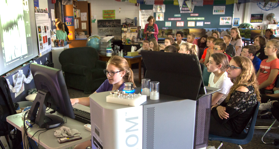
Creekside Intermediate School, Dexter, MI (June 2017)

The portable G2 Pro Phenom scanning electron microscope was set up for a demonstration of the structure of butterfly wings and how they scatter light and give the insects iridescent wings. The audience consisted of the 6th grade classes of Mrs. Denise Dutcher and Mrs. Jane Debby, 45 students in all. Students were chosen from a large number of volunteers to operate the microscope and help their classmates view the various structures of the wing colors of a Monarch butterfly.