We are excited to announce the winners of the annual (MC)2 image contest. Winners were selected by the (MC)2 scientists, who considered both creative and scientific elements of the submissions. The contest featured two categories: (1) Scanning Electron Microscopy/X-ray Microscopy/AFM and (2) Transmission Electron Microscopy.
Thank you to everyone who entered the contest for sharing your work with us!
———————————————————————————————————–
Scanning Electron Microscopy / X-ray Microscopy
1st Place
Reza Roumina, Postdoctoral Researcher, MSE, Marquis Group
“Watercolor Mosaic EBSD Map,” acquired on the TESCAN RISE SEM

The image shows the EBSD map of an additively manufactured NiCoCr alloy in the as built condition. The microstructure shows a chessboard-like grain pattern in which the boundaries contain more deformed regions at grain boundaries and crystallographic orientation gradients are observed within grains. The alloy is designed for high temperature applications requiring high creep resistance.
2nd Place
Aramanda Shanmukha Kiran, Postdoctoral Researcher, MSE, Shahani Group
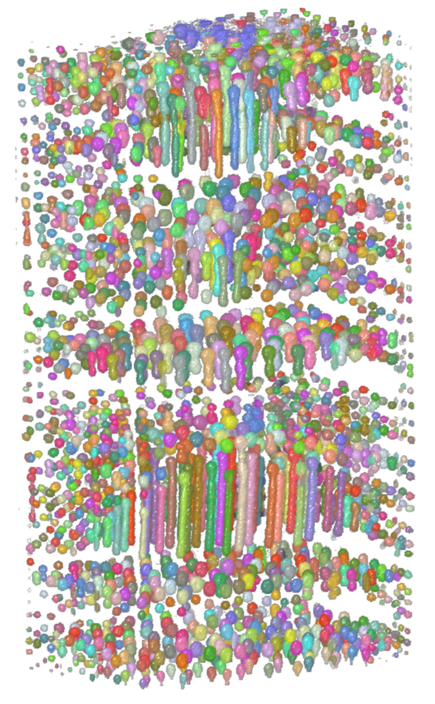
“Bismuth Droplets in Zinc Matrix,” acquired on Zeiss Xradia Versa 520 3D X-ray Microscope

This image is a microstructure of monotectic solidified sample. This is related to the study of microstructure evolution and this help to understand the mechanisms during solidification by calculating appropriate length scales in the structure. Image is colored based on the size of the particle in the data. Dragonfly is used.
Transmission Electron Microscopy
1st Place
Abby Liu, PhD Candidate, MSE, Goldman Group
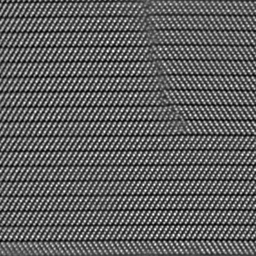
“Twin in (BiSb)2Te3” acquired on Thermo Fisher Spectra STEM

The HAADF image reveal multiple quintuple layers of (BiSb)2Te3 separated by van der Waals gaps, viewed along the [11-20]. Each quintuple layer consists of five atoms. Two different twin boundaries are present in the image, basal twin and 60° twin. At a basal twin boundary, two adjacent QLs are rotated by 180° along the c-axis, resulting in a mirror plane parallel to the surface. On the other hand, at a 60° twin boundary, the mirror plane is perpendicular to the surface, and the atomic structures within the QLs are mirrored across the basal plane.
2nd Place
Mackenzie Warwick, PhD Candidate, NERS, Field Group
“Let’s get creepy: an investigation of multi-length-scale irradiation-induced defects under irradiation creep conditions,” acquired on the Thermo Fisher Tecnai G2 F30 TWIN

My dissertation work focuses on the low nucleation dose, high temperature regime of irradiation creep and how the radiation-induced defect clusters of interstitials and vacancies impact the motion (climb-glide), orientation, and growth of dislocation lines. This novel stress-gradient specimen geometry was irradiated to 0.2 dpa at a temperature of 550C, and this was a targeted (100) grain lift-out taken -15 degrees perpendicular to the applied stress of 237 MPa. The larger micrograph shows a cellular structure of dislocations, which is common when one of the <100> planes are aligned with the applied stress. The smaller inset micrograph shows incredibly tiny defect clusters with an average diameter of 7.45 nm, also with the same preferential alignment as the large dislocation lines. These results are particularly important as current irradiation creep theory excludes small defect clusters and their impact(s) on dislocation pinning, radiation induced segregation, and their contribution to “negative” strain observed during irradiation. Previous lamella preparation techniques and TEM resolution has not allowed us to discern between FIB-induced damage and real defect clusters.
